


Kidney stone
A stone may form due to crystallization of lithogenic factors in the upper urinary tract, it can subsequently move into the ureter and cause renal colic. Although kidney stone is rarely fatal, patients who have had renal colic report that it is the worst pain they have ever experienced.
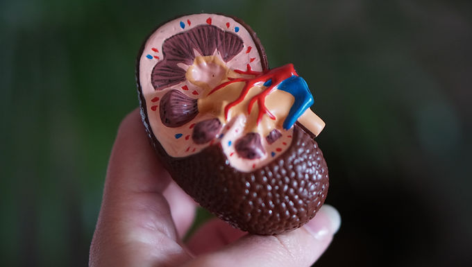
There are various types of kidney stones. It is clinically important to identify the stone type, which informs prognosis and selection of the optimal preventive regimen. Calcium oxalate stones are most common (~75%); next, in order, are calcium phosphate (~15%), uric acid (~8%), struvite (~1%), and cystine (<1%) stones. Many stones are a mixture of crystal types (e.g., calcium oxalate and calcium phosphate) and also contain protein in the stone matrix. Rarely, stones are composed of medications, such as acyclovir, indinavir, and triamterene. Infectious stones, if not appropriately treated, can have devastating consequences and lead to end-stage renal disease.
Nephrolithiasis is a global disease likely due to Westernization of lifestyle habits (e.g., dietary changes, increasing body mass index).Once an individual has had a stone, the prevention of a recurrence is essential. Most experts agree that radiographic evidence of a second stone should be considered to represent a recurrence, even if the stone has not yet caused symptoms.
Risk factors for nephrolithiasis can be categorized as dietary, nondietary, or urinary. Efforts to avoid high oxalate intake, higher intake of animal protein, higher sodium and sucrose intake should be beneficial. Avoidance of foods that contain high amounts of oxalate, such as spinach, rhubarb, and potatoes, is prudent. Calcium oxalate stone formers should be advised to avoid vitamin C supplements. The risk of stone formation increases as urine volume decreases. When the urine output is less than 1 L/d, the risk of stone formation more than doubles. Fluid intake is the main determinant of urine volume. Coffee, tea, beer, and wine are associated with a reduced risk of stone formation. Sugar-sweetened carbonated beverage consumption may increase risk.
The incidence of stone disease is highest in middle-aged white men, but stones can form in infants as well as in the elderly. Weight gain increases the risk of stone formation, Environmental and occupational influences that may lead to lower urine volume, such as working in a hot environment or lack of ready access to water or a bathroom, are important considerations.
Urine pH influences the solubility of some crystal types. Uric acid stones form only when the urine pH is consistently ≤5.5 or lower, whereas calcium phosphate stones are more likely to form when the urine pH is ≥6.5 or higher. Cystine is more soluble at higher urine pH. Calcium oxalate stones are not influenced by urine pH.
The risk of nephrolithiasis is more than twofold greater in individuals with a family history of stone disease. This association is likely due to a combination of genetic predisposition and similar environmental exposures.
It typically requires weeks to months (and often much longer) for a kidney stone to grow to a clinically detectable size. Although the passage of a stone is a dramatic event, stone formation and growth are characteristically clinically silent. A stone can remain asymptomatic in the kidney for years or even decades before signs (e.g., hematuria) or symptoms (e.g., pain) become apparent. Thus, it is important to remember that the onset of symptoms, typically attributable to a stone moving into the ureter, does not provide insight into when the stone actually formed.
There are two common presentations for individuals with an acute stone event: renal colic and painless gross hematuria.
Renal colic is a misnomer because pain typically does not subside completely; rather, it varies in intensity. When a stone moves into the ureter, the discomfort often begins with a sudden onset of unilateral flank pain. The intensity of the pain can increase rapidly, and there are no alleviating factors. This pain, which is accompanied often by nausea and occasionally by vomiting, may radiate, depending on the location of the stone. If the stone lodges in the upper part of the ureter, pain may radiate anteriorly; if the stone is in the lower part of the ureter, pain can radiate to the ipsilateral testicle in men or the ipsilateral labium in women. Occasionally, a patient has gross hematuria without pain.
The urine will contain red and white blood cells, but the urine culture will be negative. An obstructing stone with proximal infection may present as acute pyelonephritis.
The diagnosis is often made on the basis of the history, physical examination, and urinalysis. Thus, it may not be necessary to wait for radiographic confirmation before treating the symptoms. Helical CT detects stones as small as 1 mm that may be missed by other imaging modalities. Typically, helical CT reveals a ureteral stone or evidence of recent passage (e.g., perinephric stranding or hydronephrosis), whereas a plain abdominal radiograph (kidney/ureter/bladder, or KUB) can miss a stone in the ureter or kidney, even if it is radiopaque, and does not provide information on obstruction. Abdominal ultrasound offers the advantage of avoiding radiation and provides information on hydronephrosis, but it is not as sensitive as CT and images only the kidney and possibly the proximal segment of the ureter; thus most ureteral stones are not detectable by ultrasound. Excessive fluid administration has not been shown to be beneficial; therefore, the goal should be to maintain euvolemia.
The urine volume should be at least 2 L/d. Because of differences in insensible fluid losses and fluid intake from food sources, the required total fluid intake will vary from person to person. Rather than specify how much to drink, it is more helpful to educate patients about how much more they need to drink in light of their 24-h urine volume. For example, if the daily urine volume is 1.5 L, then the patient should be advised to drink at least 0.5 L more per day in order to increase the urine volume to the goal of 2 L/day.
Is homeopathy good for kidney stone?
Answer
Homeopathy is an ideal and effective mode of treatment for kidney stone. It dissolve the stone smoothly without any need for surgery.
How long does it take homeopathy to cure kidney stones?
Answer
Homeopathic treatment flushes out kidney stone with a month whether its small or big
What management required for kidney stone?
Answer
Drink plenty of fluids, especially water